चित्र:Rheumatoid arthritis ultrasound MRI MCP joint ar1904-2.gif
नेविगेशन पर जाएँ
खोज पर जाएँ


इस पूर्वावलोकन का आकार: ४१४ × ५९९ पिक्सेल । अन्य resolutions: १६६ × २४० पिक्सेल | ३३२ × ४८० पिक्सेल | १,००४ × १,४५२ पिक्सेल ।
मूल फ़ाइल (१,००४ × १,४५२ चित्रतत्व, संचिका का आकार: ७९५ KB, माइम प्रकार: image/gif)
| विवरण |
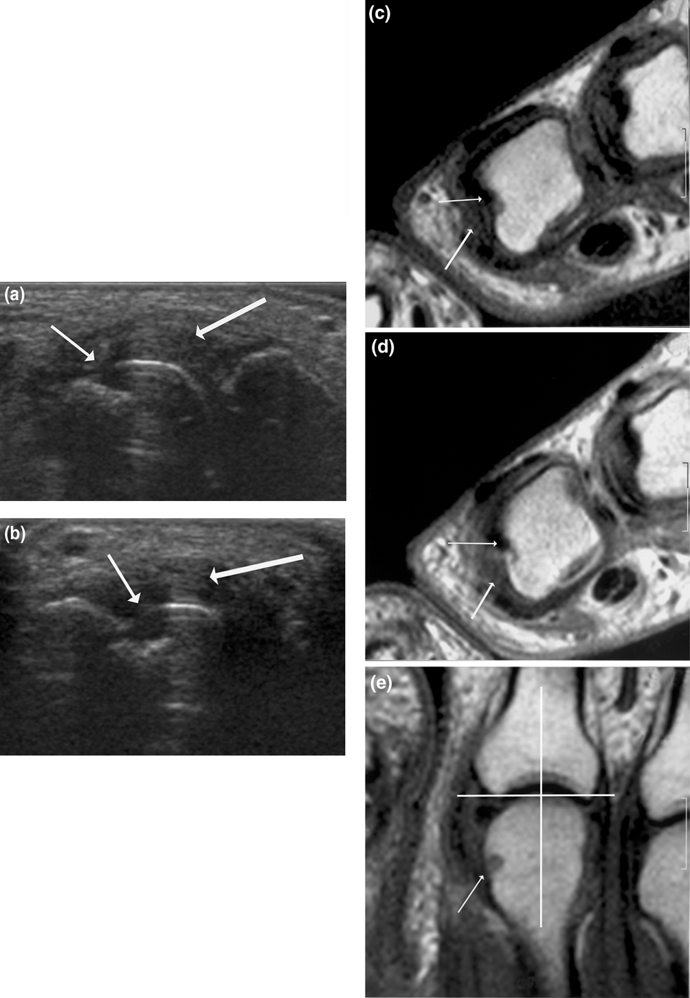
English: Signs of destruction and inflammation on ultrasonography and MRI in second metacarpophalangeal joint: established RA. Thin arrows indicate an erosive change; thick arrows indicate synovitis. Ultrasonography in the (a) longitudinal and (b) the transverse planes shows both signs of destruction (grade 2) and inflammation (grade 3). Axial T1-weighted magnetic resonance images were obtained (c) before and (d) after contrast administration (grade 3 synovitis). Additionally, a coronal T1-weighted magnetic resonance image (e) before contrast administration visualizes the same bone erosion as shown in panels c and d. The coronal magnetic resonance image of the second metacarpophalangeal joint (panel e) is additionally covered by a grid illustrating division of the assessed joints into quadrants: proximal radial, proximal ulnar, distal radial and distal ulnar. MRI, magnetic resonance imaging; RA, rheumatoid arthritis. |
| दिनांक | Published: 6 March 2006 |
| स्रोत | Ultrasonography of the metacarpophalangeal and proximal interphalangeal joints in rheumatoid arthritis: a comparison with magnetic resonance imaging, conventional radiography and clinical examination. Arthritis Research & Therapy 2006, 8:R52. doi:10.1186/ar1904 |
| लेखक | Marcin Szkudlarek, Mette Klarlund, Eva Narvestad, Michel Court-Payen, Charlotte Strandberg, Karl E Jensen, Henrik S Thomsen and Mikkel Østergaard. |
लाइसेंस
इस फ़ाइल को क्रिएटिव कॉमन्स श्रेय 2.0 साधारण लाइसेंस के अंतर्गत लाइसेंस किया गया है।
- आप खुलकर:
- बाँट सकते हैं – रचना की प्रतिलिपि बना सकते हैं, बाँँट सकते हैं और संचारित कर सकते हैं
- रीमिक्स कर सकते हैं – कार्य को अनुकूलित कर सकते हैं
- निम्नलिखित शर्तों के अंतर्गत:
- श्रेय – यह अनिवार्य है कि आप यथोचित श्रेय प्रदान करें, लाइसेंस की कड़ी प्रदान करें, और अगर कोई बदलाव हुए हों तो उन्हें इंगित करें। आप ऐसा किसी भी उचित तरीके से कर सकते हैं, लेकिन किसी भी तरह उससे यह नहीं संकेत नहीं किया जाना चाहिए कि लाइसेंसधारी द्वारा आपको अथवा आपके इस प्रयोग का समर्थन किया जा रहा हो।
फ़ाइल का इतिहास
फ़ाइल पुराने समय में कैसी दिखती थी यह जानने के लिए वांछित दिनांक/समय पर क्लिक करें।
| दिनांक/समय | अंगूठाकार प्रारूप | आकार | प्रयोक्ता | टिप्पणी | |
|---|---|---|---|---|---|
| वर्तमान | १७:०९, ७ फ़रवरी २००९ | | १,००४ × १,४५२ (७९५ KB) | wikimediacommons>Stevenfruitsmaak | {{Information |Description={{en|1=Signs of destruction and inflammation on ultrasonography and MRI in second metacarpophalangeal joint: established RA. Thin arrows indicate an erosive change; thick arrows indicate synovitis. Ultrasonography in the (a) lon |
फ़ाइल का उपयोग
इस फ़ाइल का प्रयोग नीचे दिए गए पृष्ठ पर हो रहा है: